
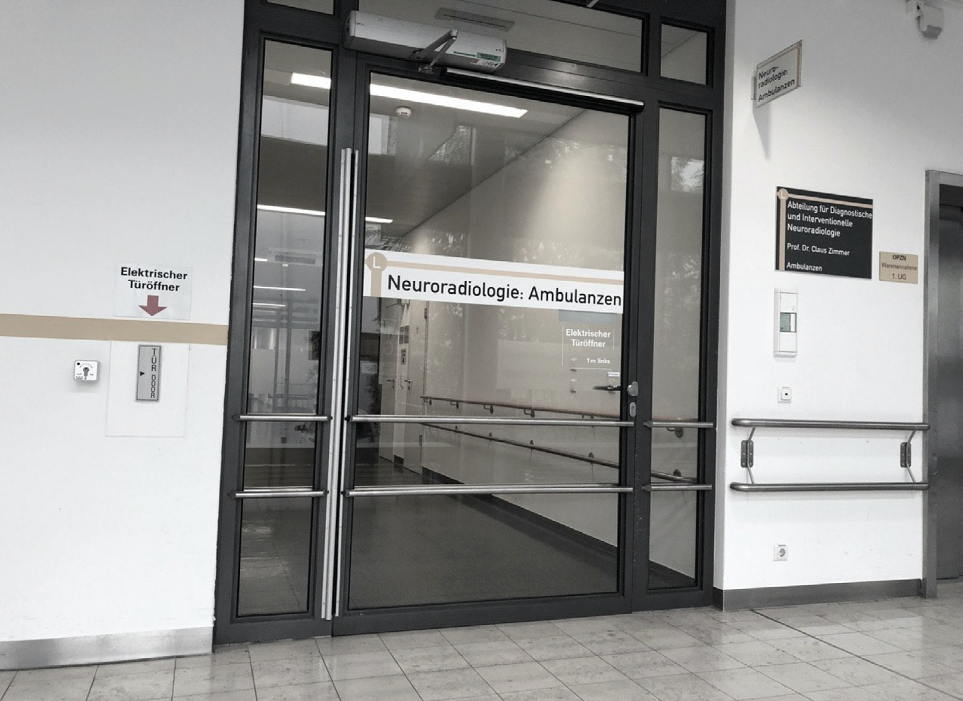
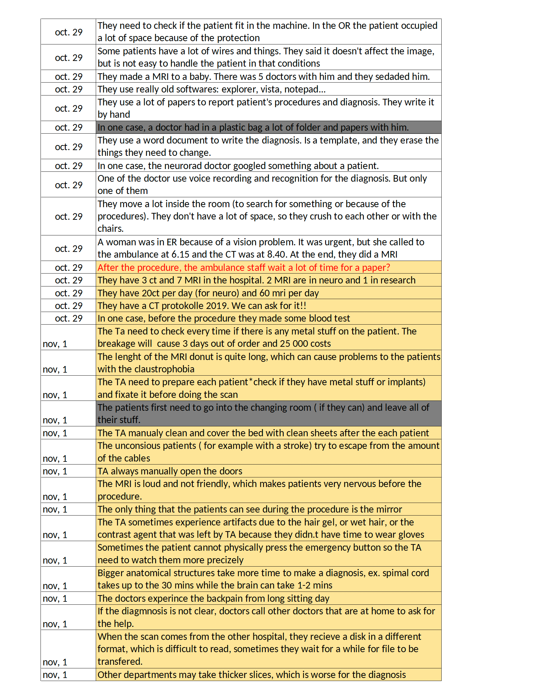
Part of the observation documents
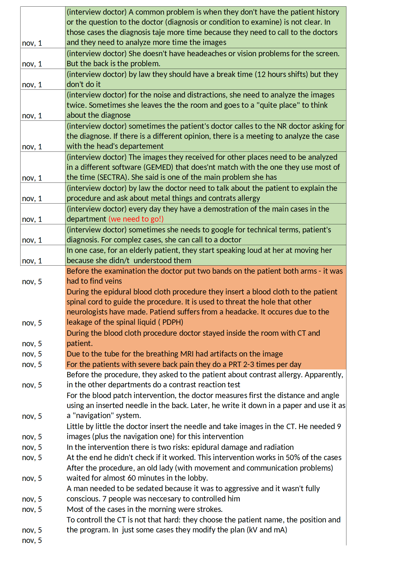
Part of the observation documents
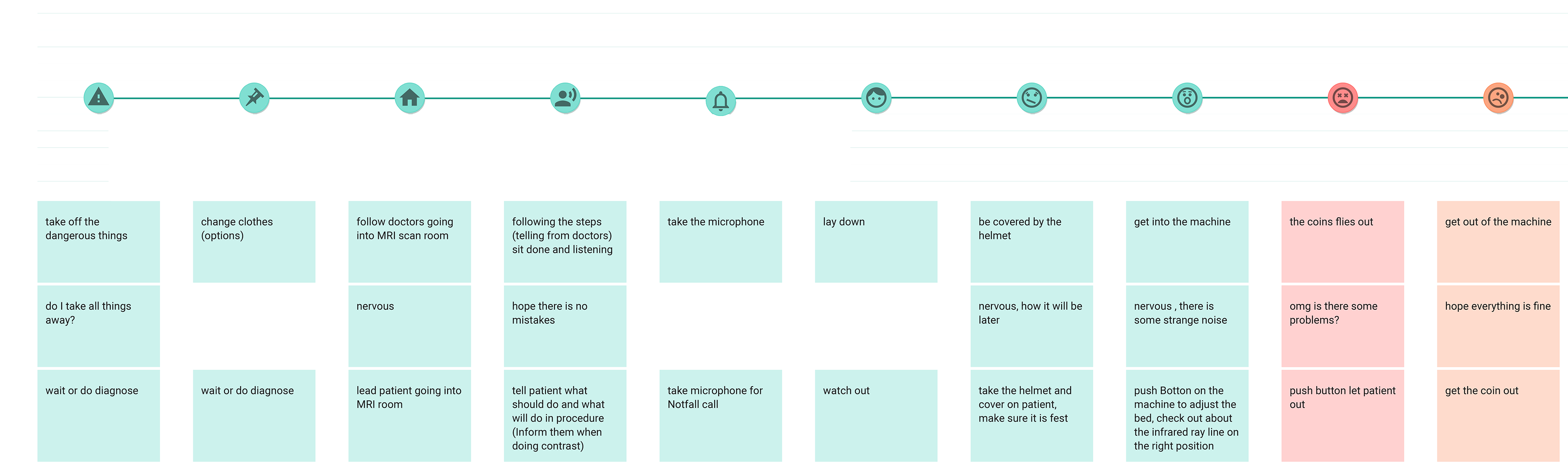
User journey in the clinic from the observation and research
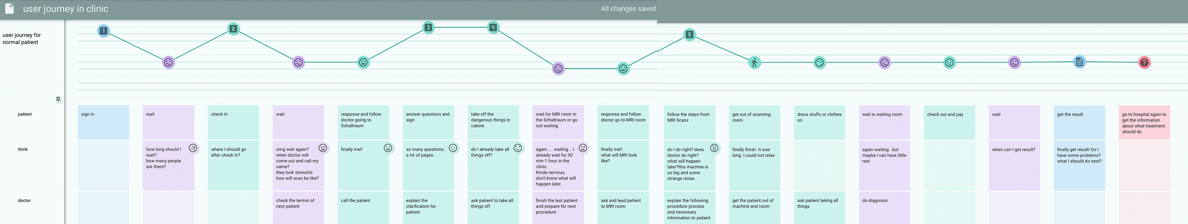
User journey in the clinic from the observation and research
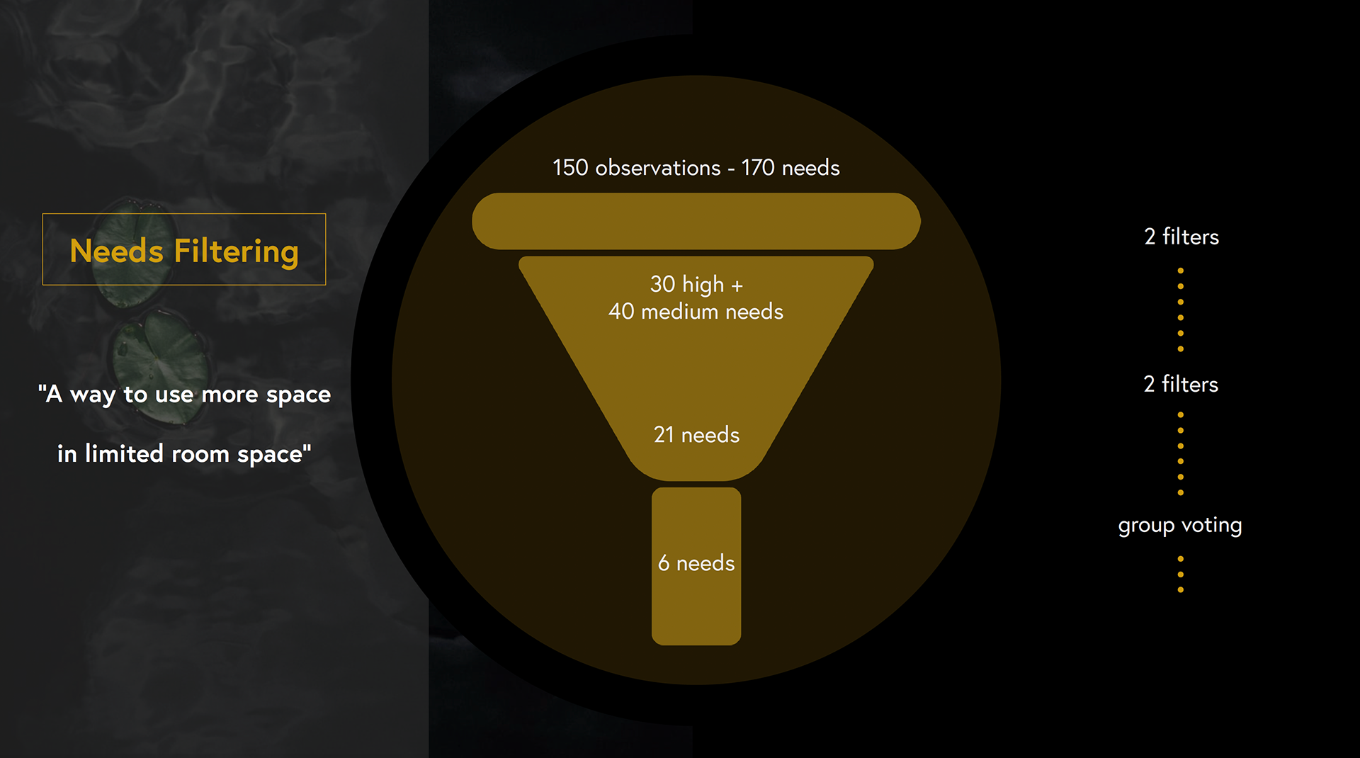
How we filtered the 170 needs down to 6 needs
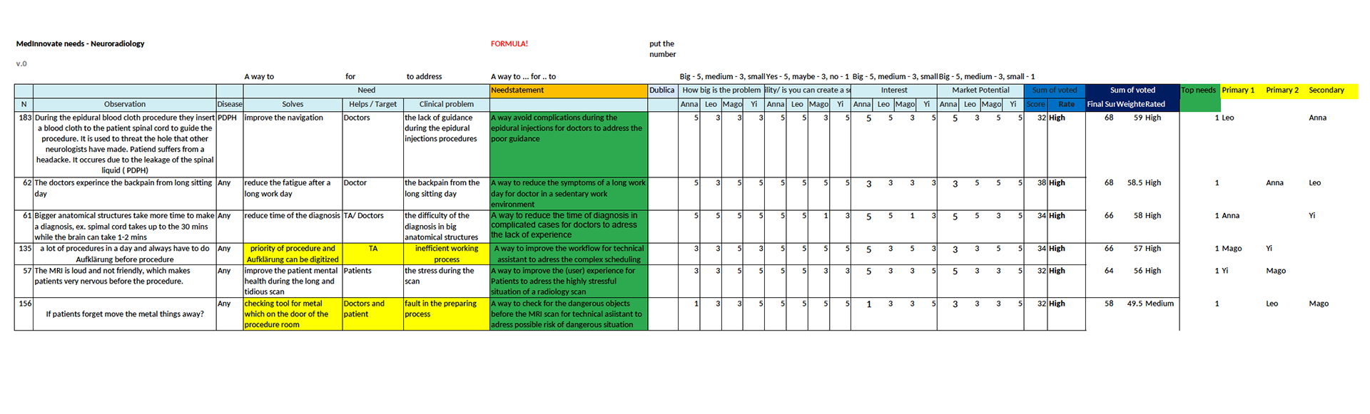
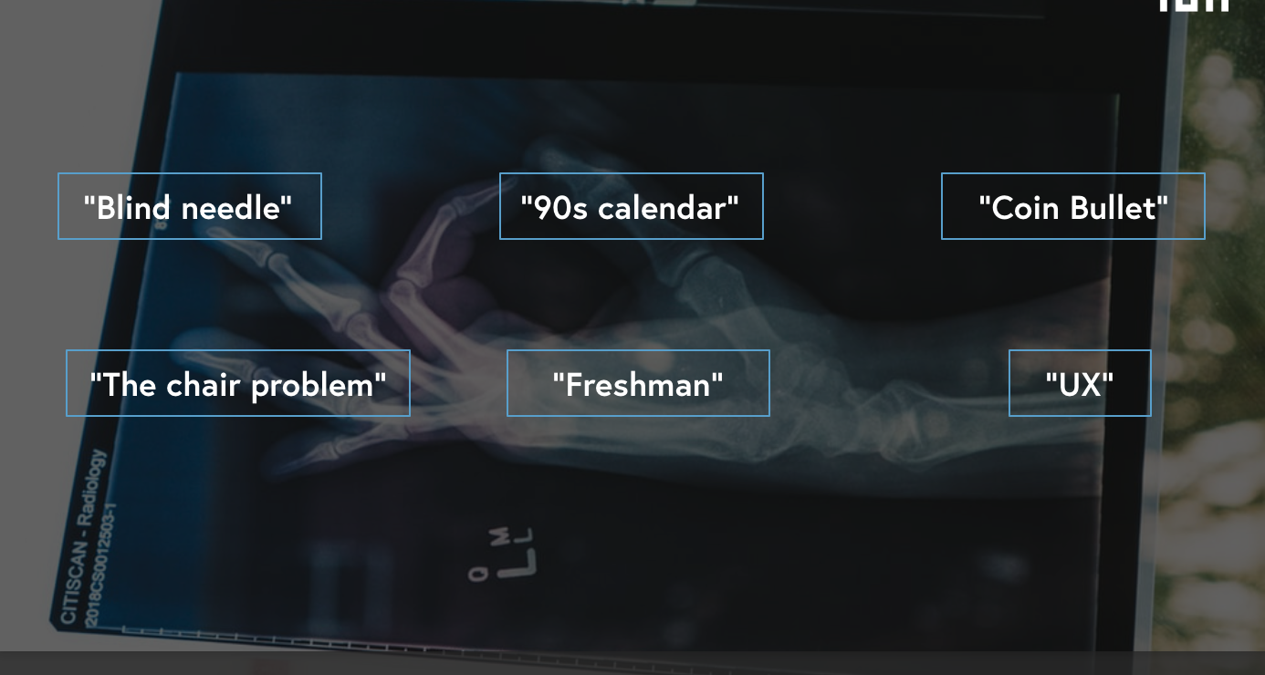
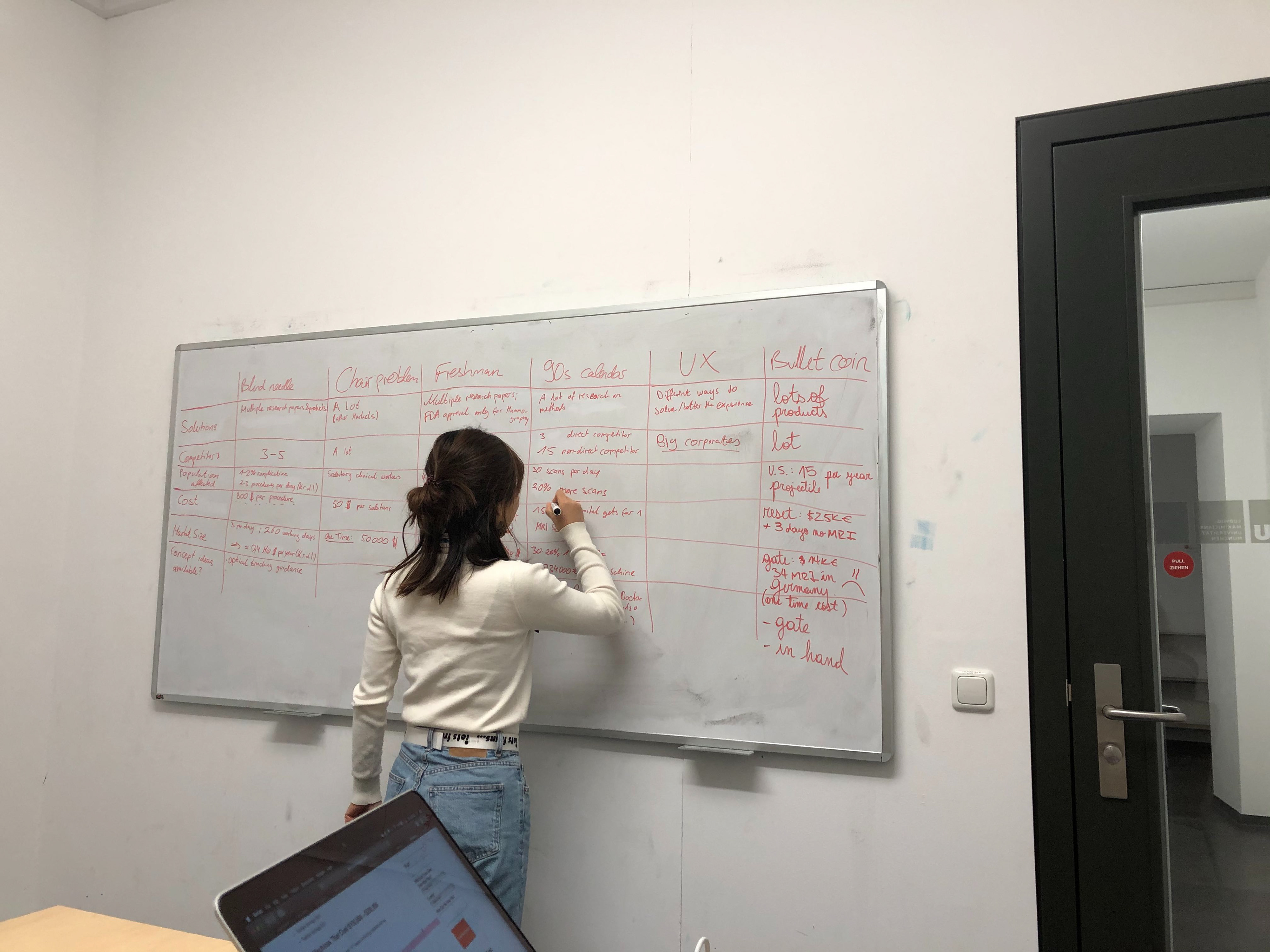
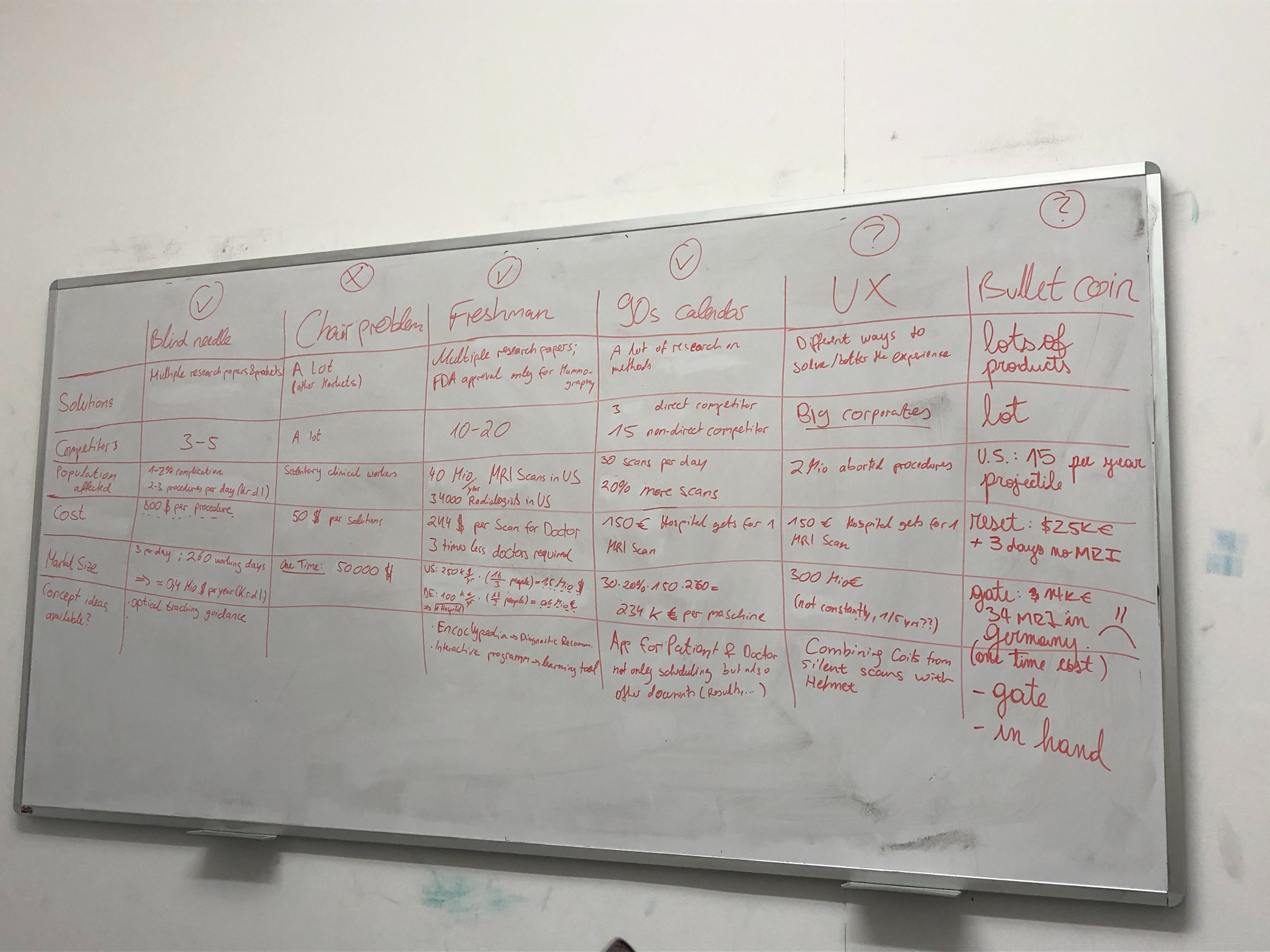
Part of the filtering process: how we filter and narrow down the problems and needs
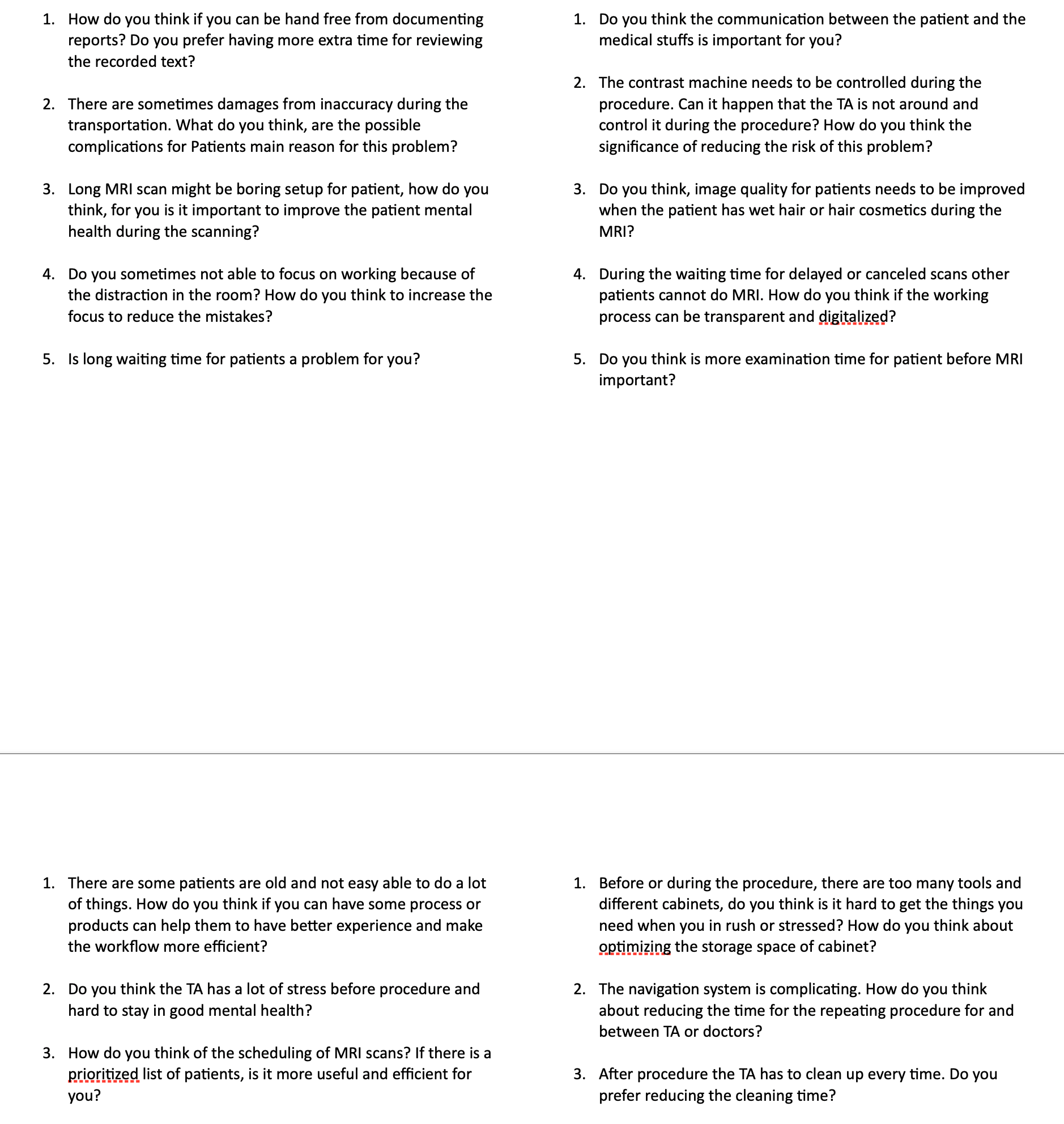
Interview questions
6 Needs
Me writing down the criteria for filtering secondary needs
Secondary needs filtering
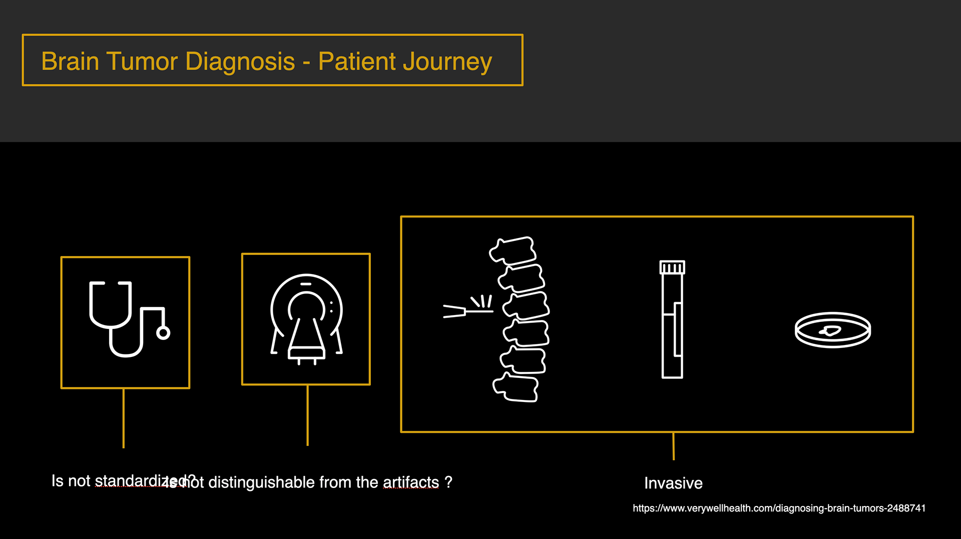
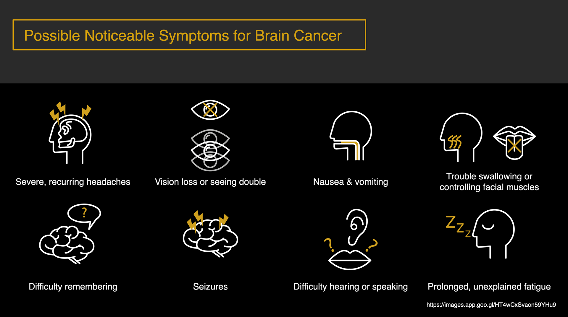
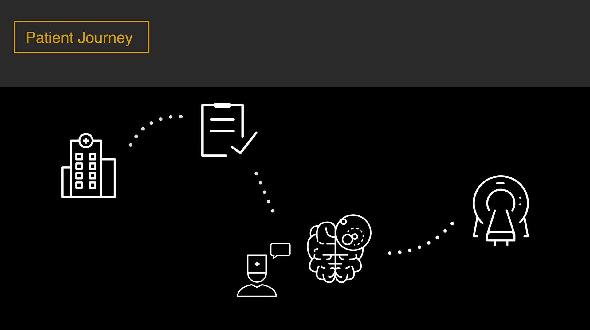
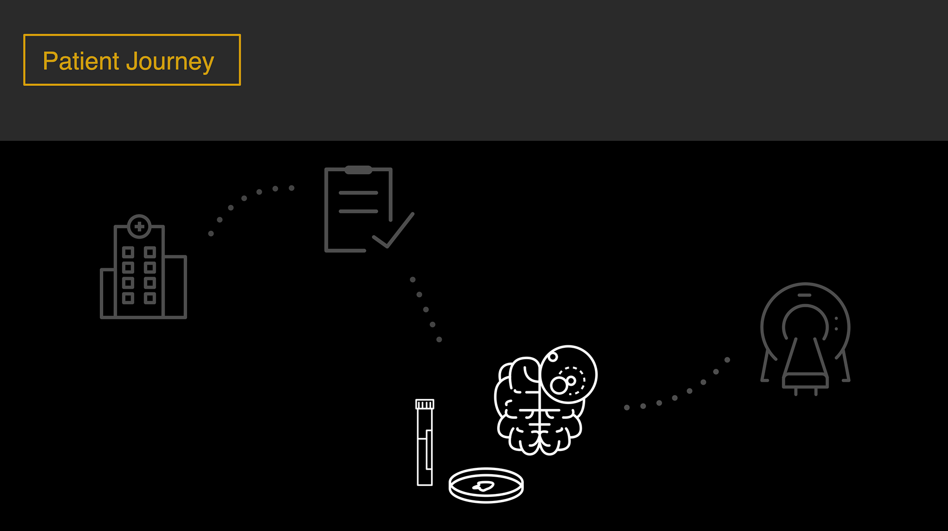
Patient journey for the case of brain tumor diagnosis and its problems
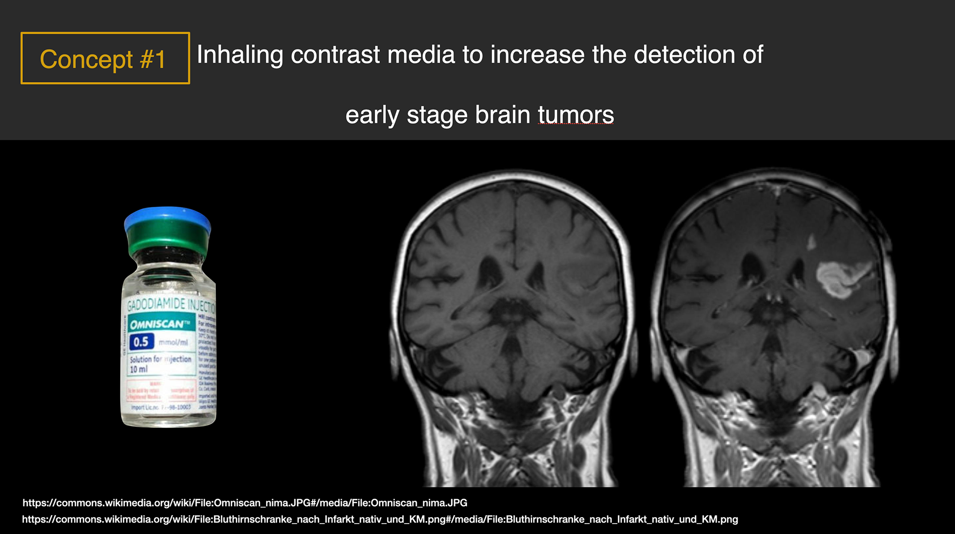
Concept 1
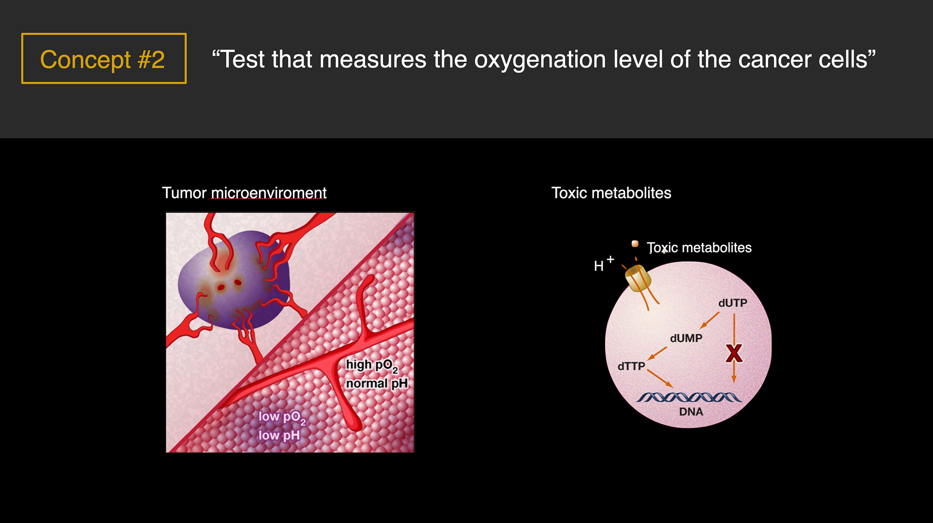
Concept 2
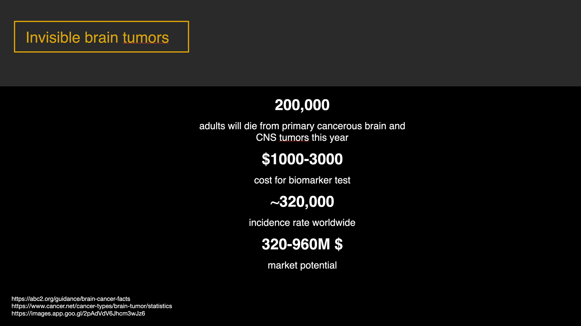
Market potential
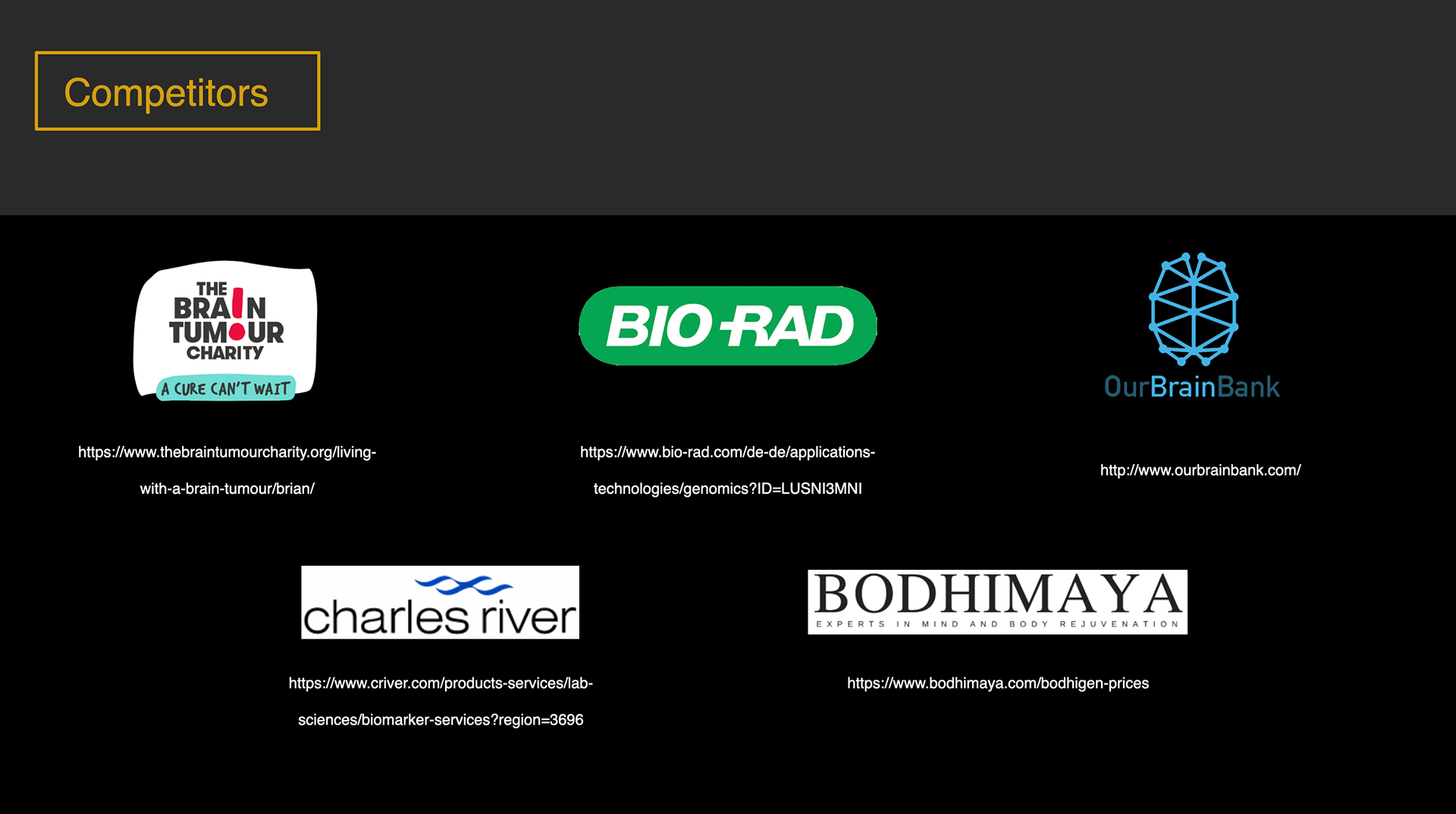
Competitors
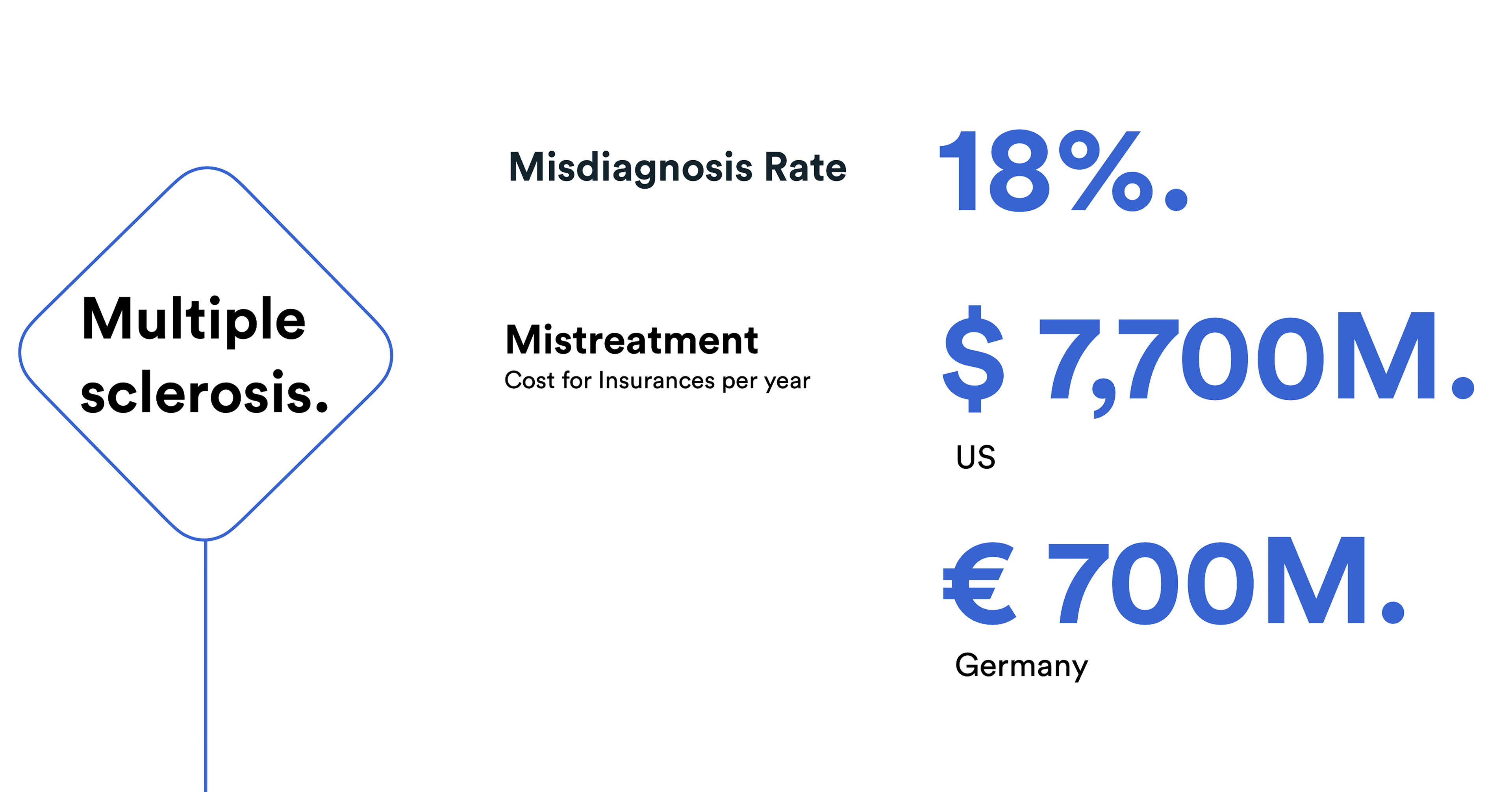
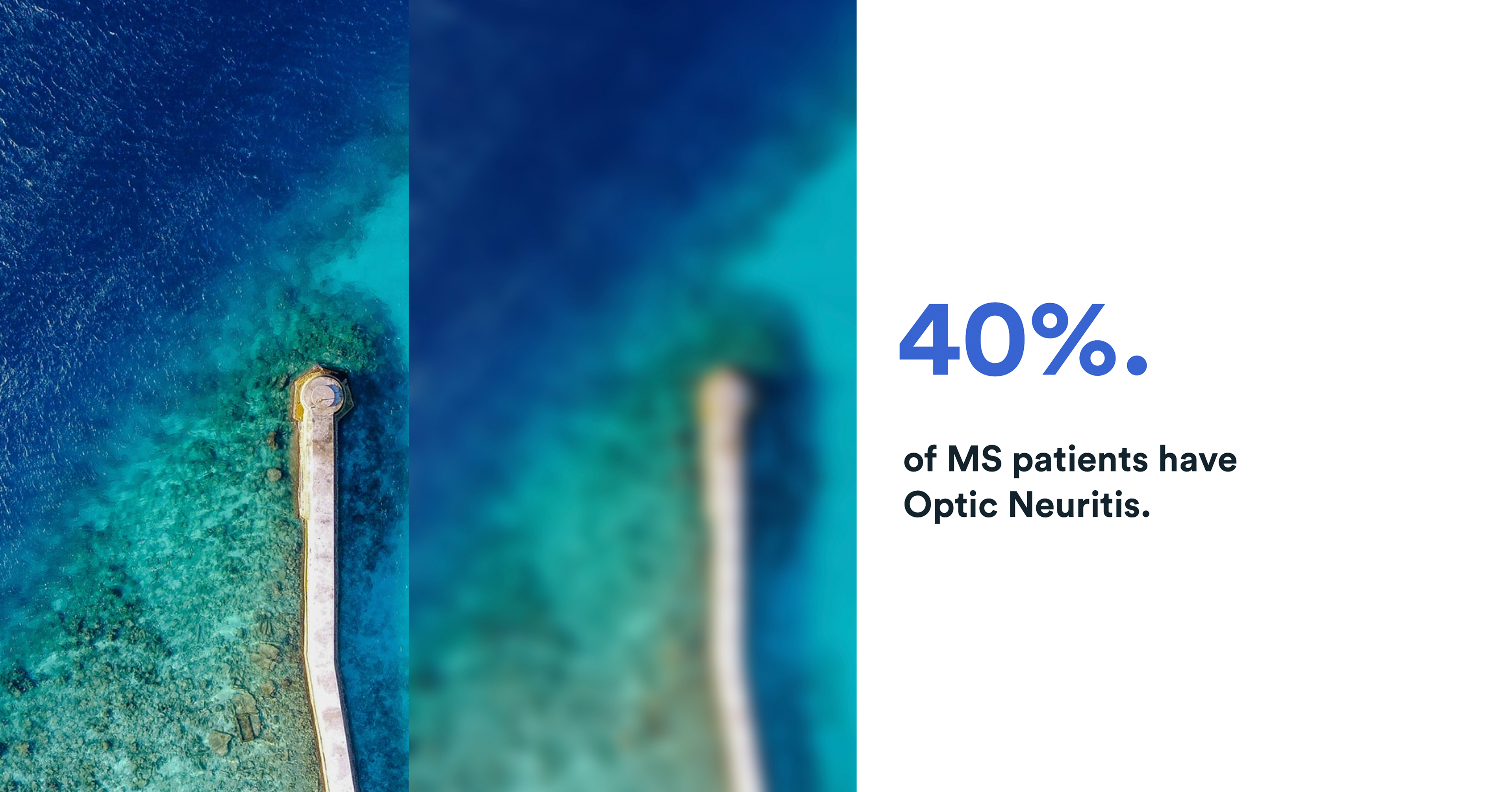
Possible noticeable symptoms for multiple sclerosis
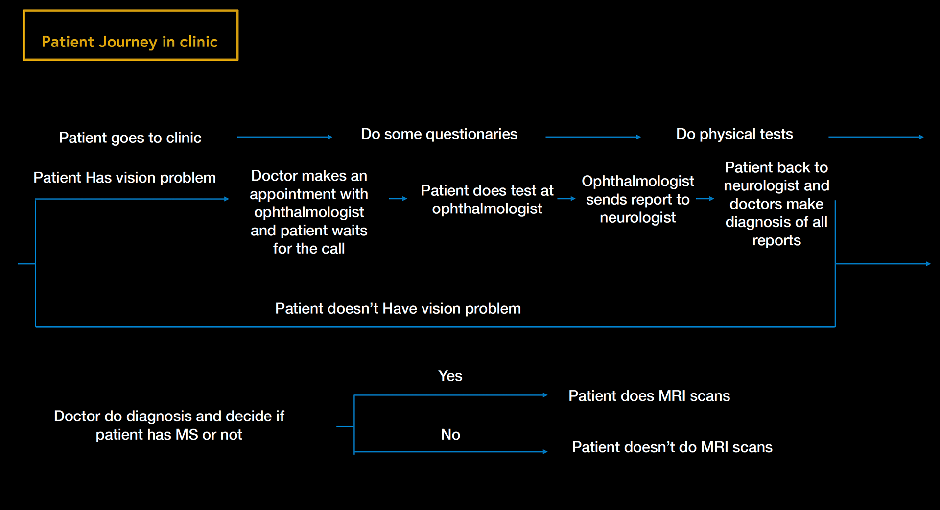
Patient journey for MS patient in clinic
Change the current solution to the quantitive objective tests
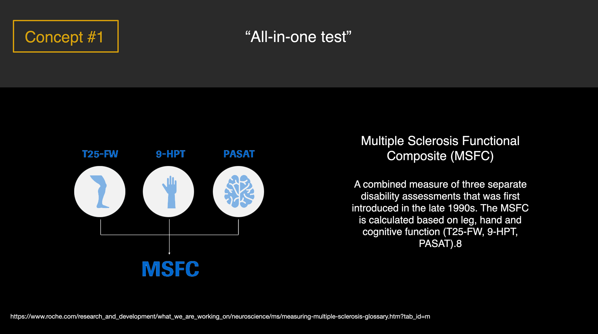
Concept 1
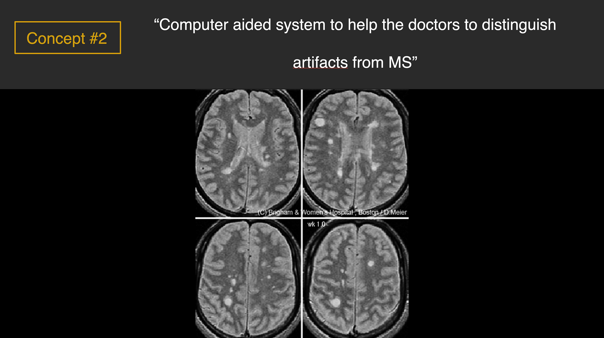
Concept 2
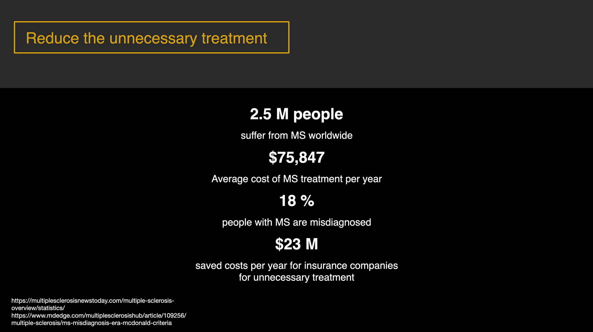
Market potential
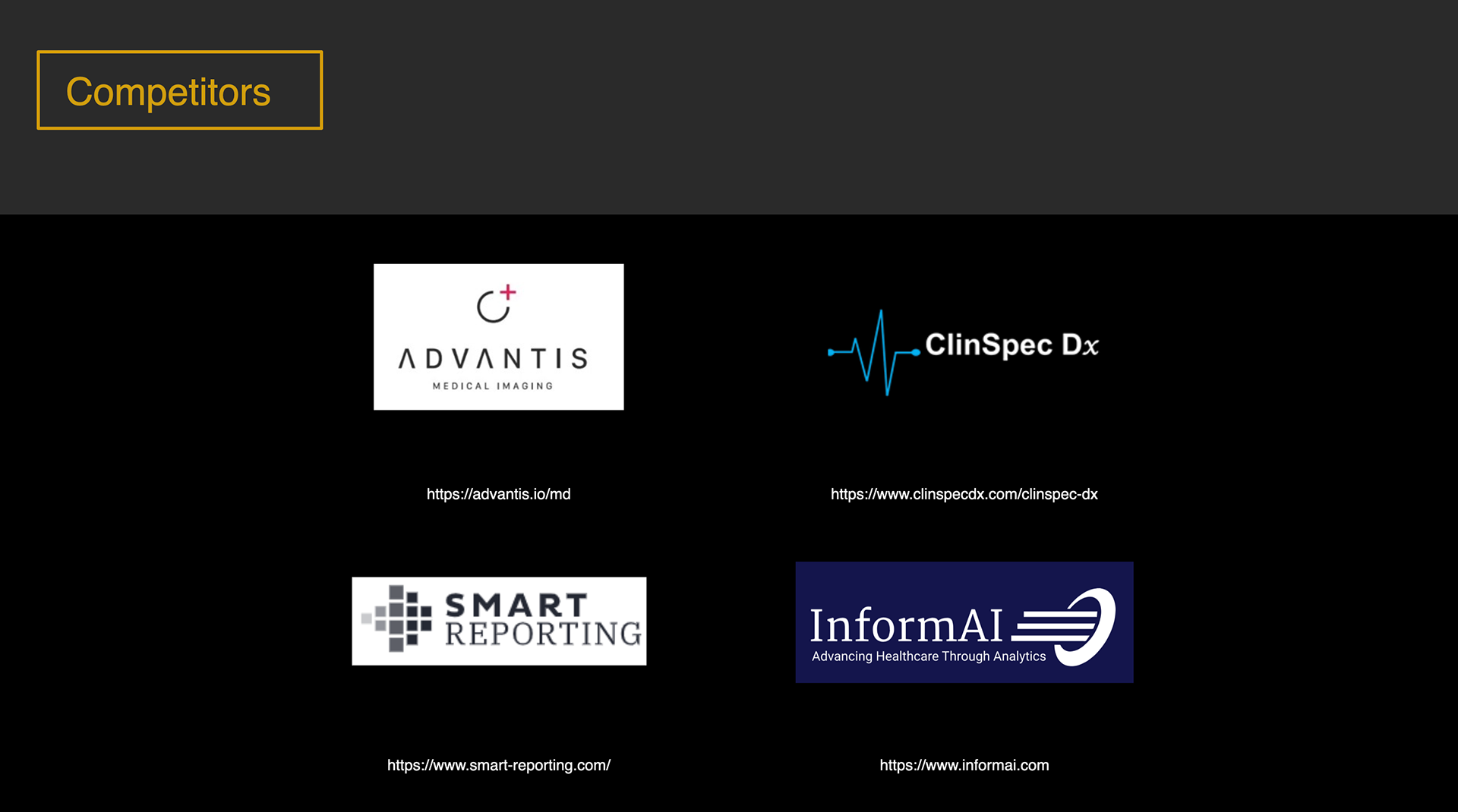
Competitors

Sketch concept of MS visual test device
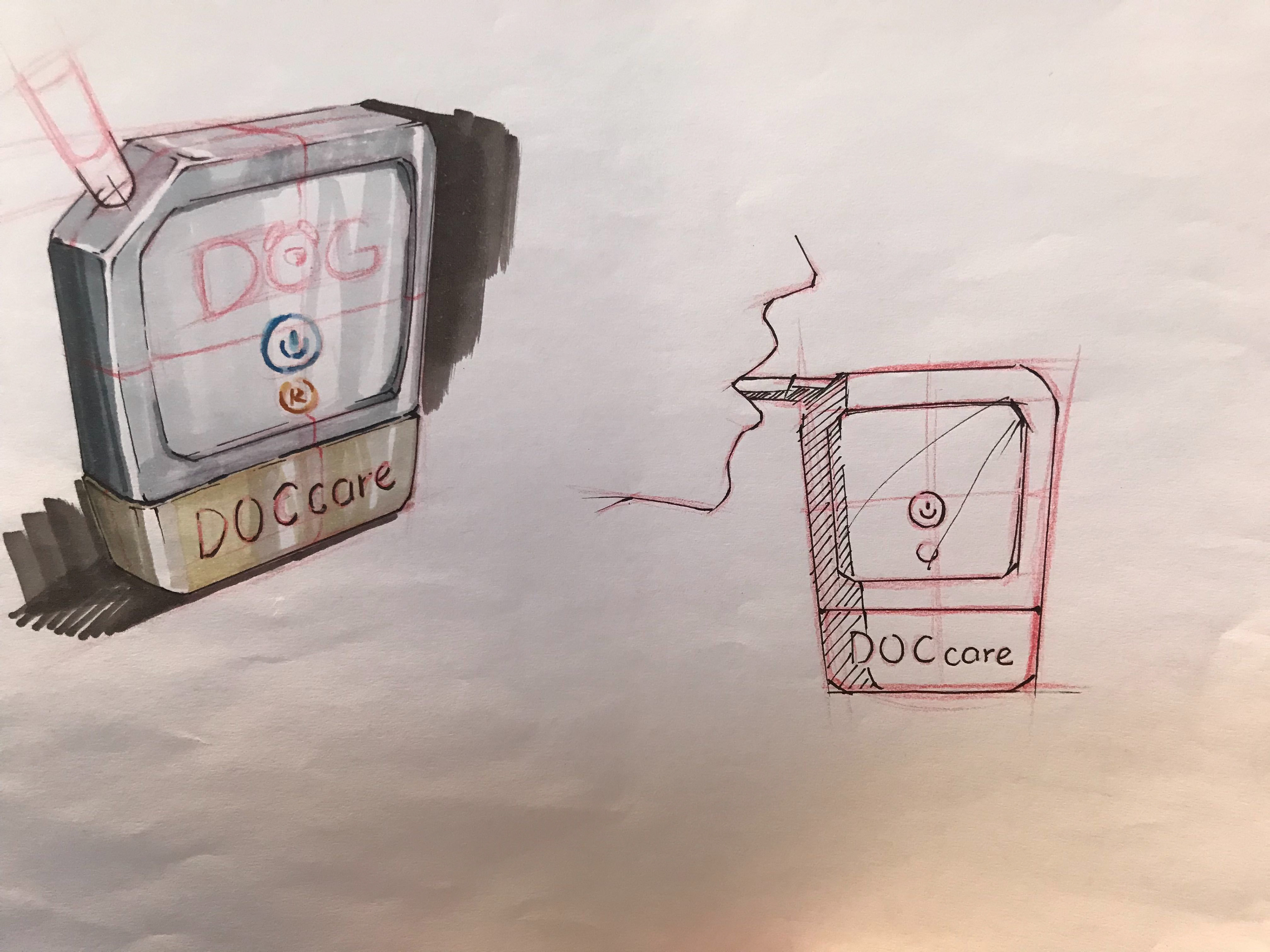
Sketch concept of the brain tumor measuring breathalyzer

First design concept statement
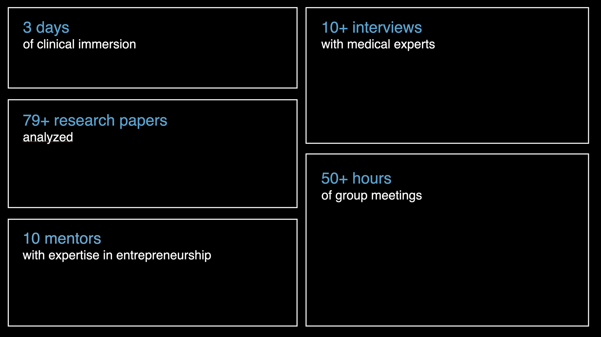
How did we get there
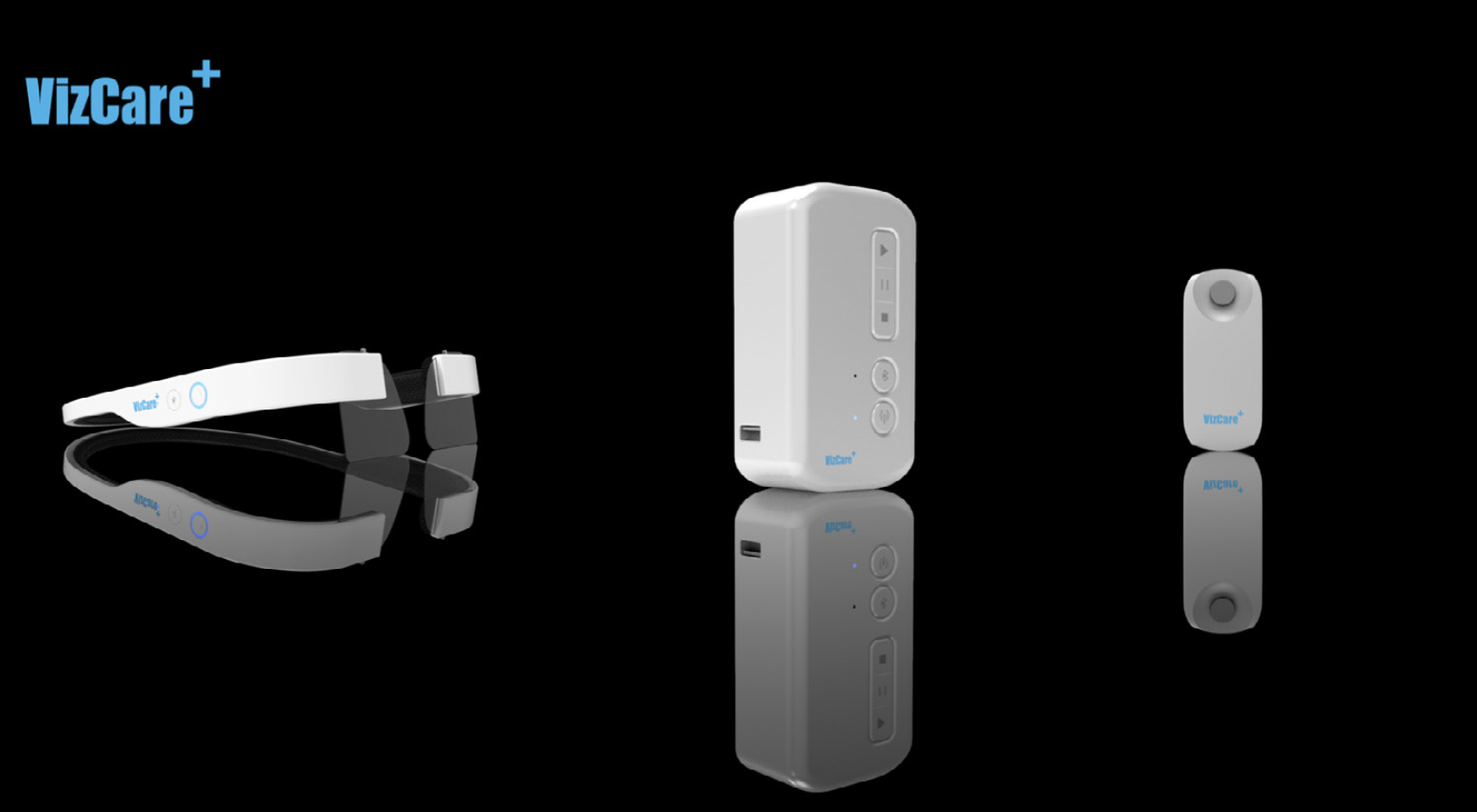
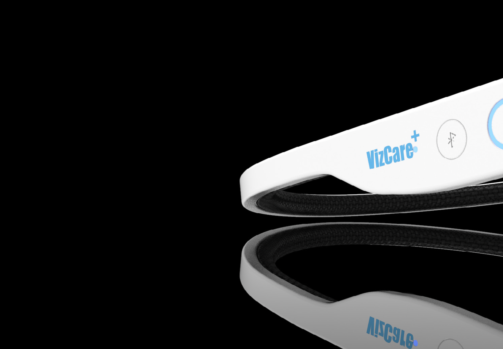
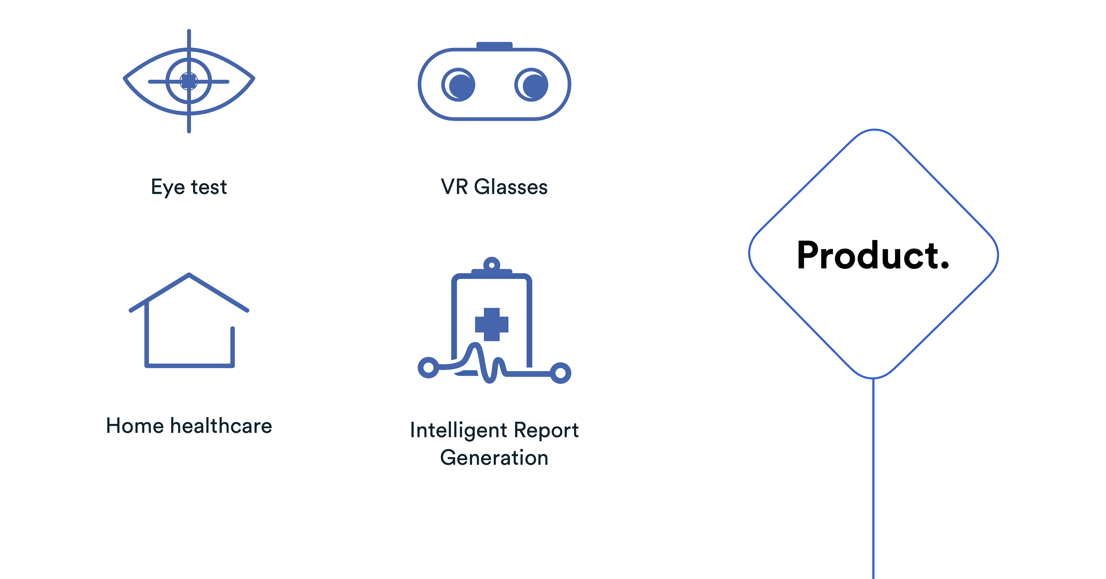
Physical products
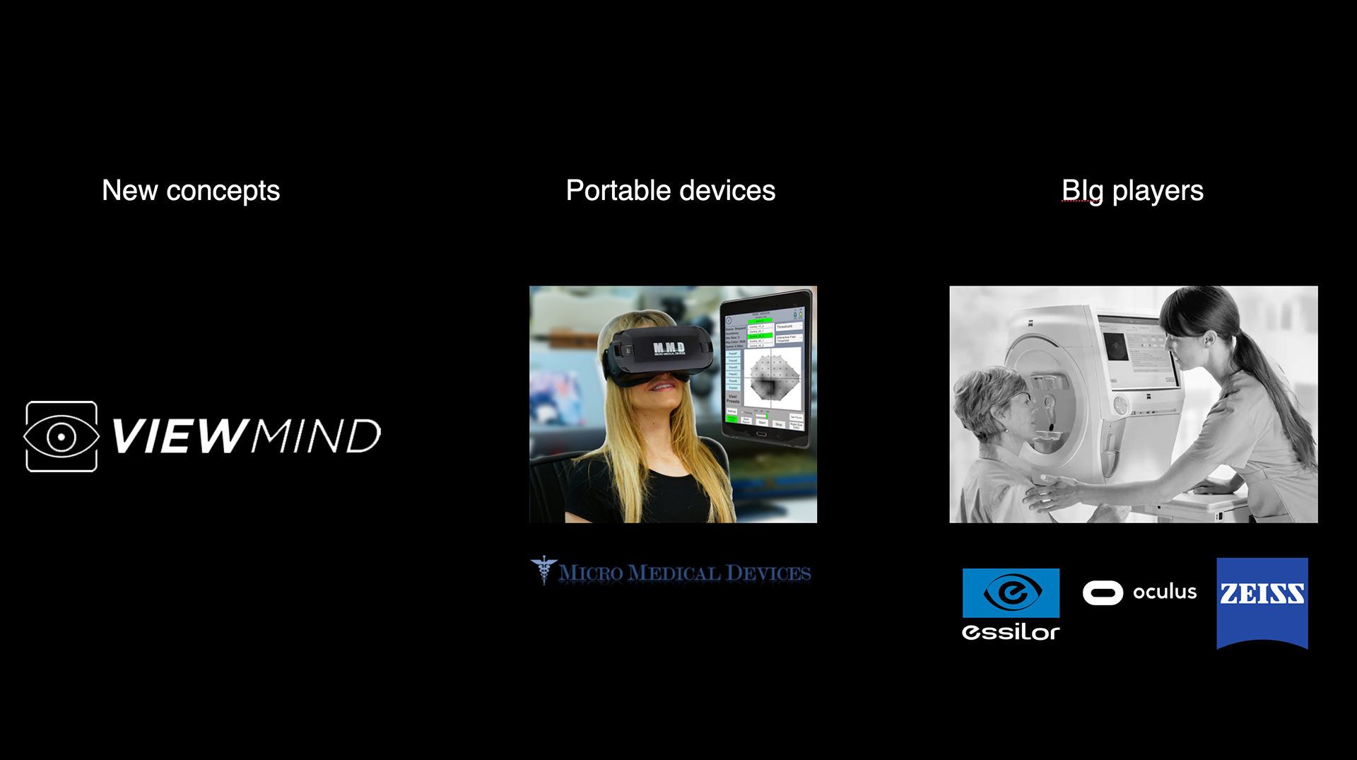
Competitors
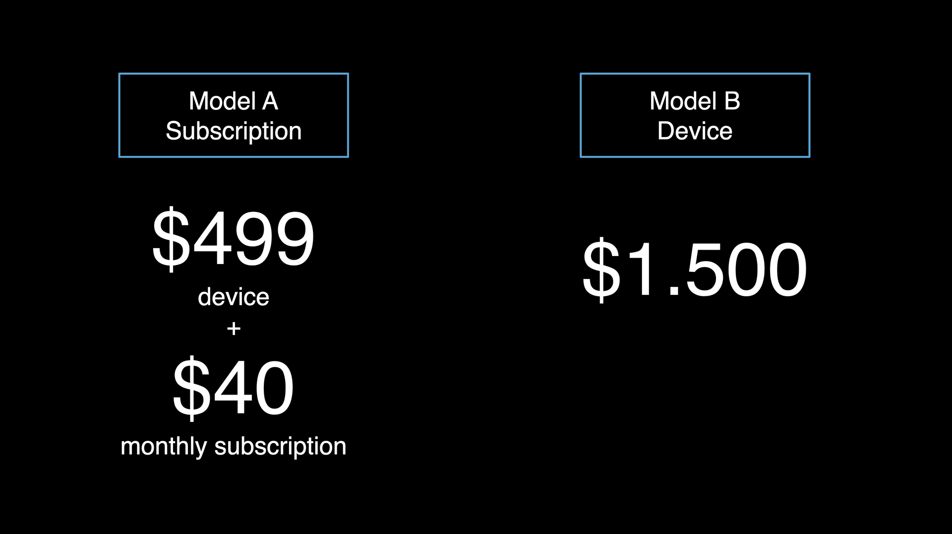
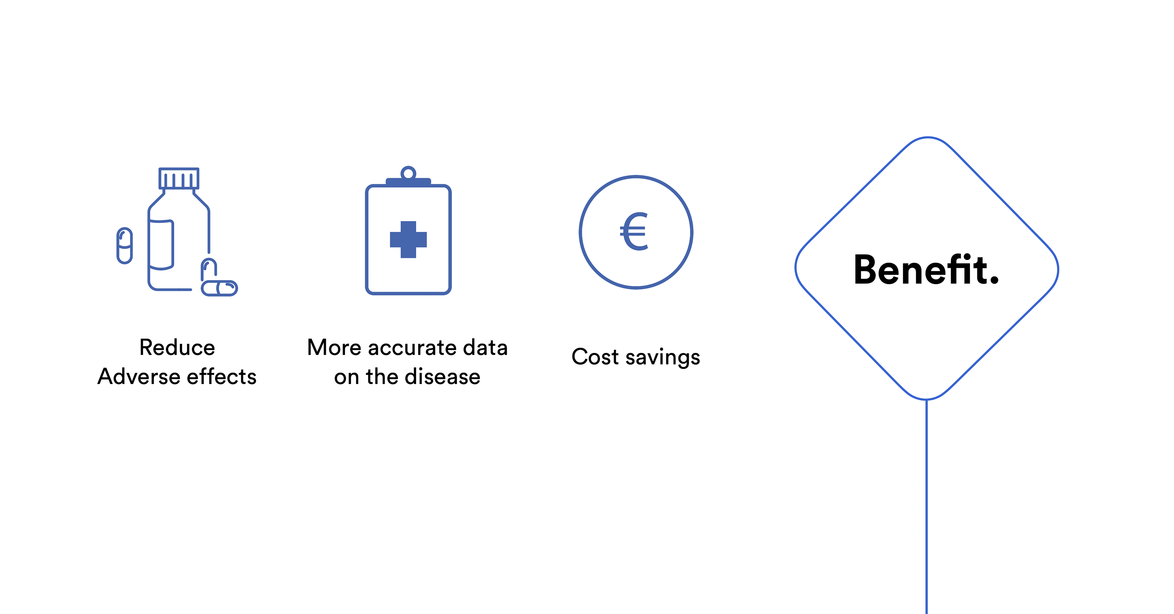
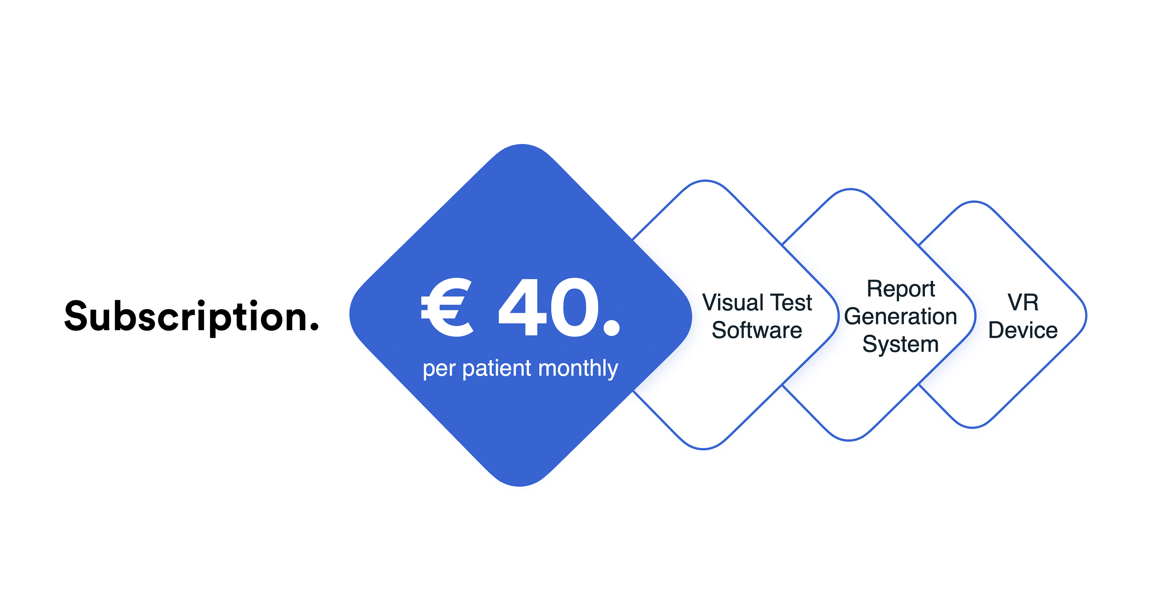
Business model
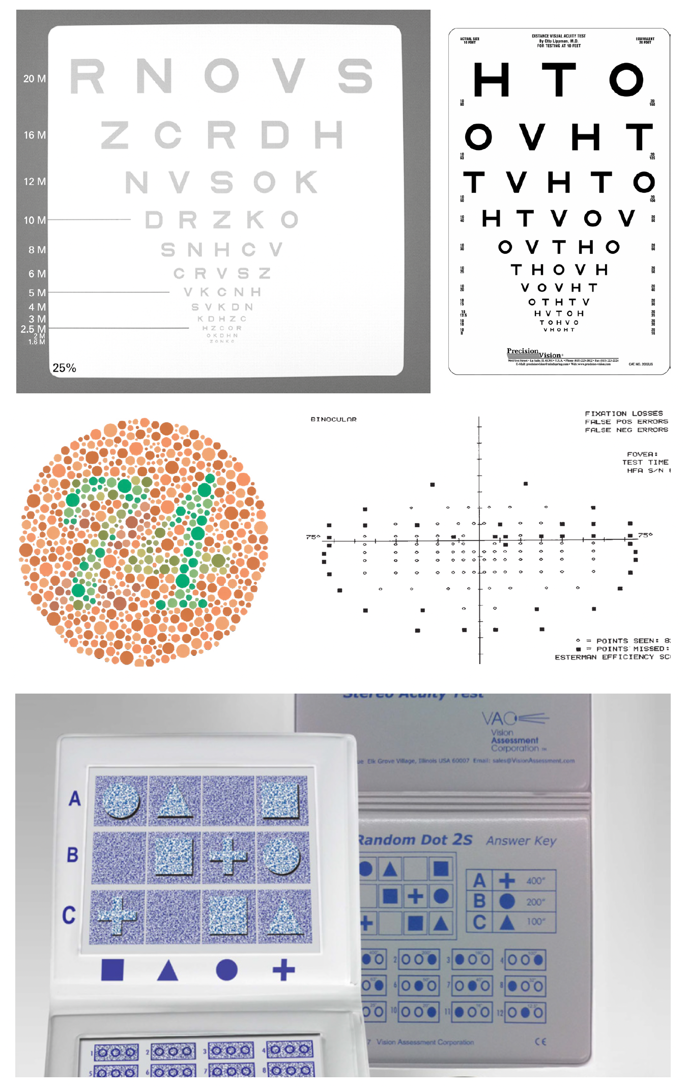
Visual example of low contrast, visual acuity, color vision, visual field, and depth perception
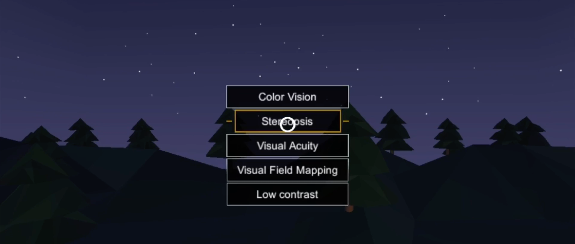
Eye tests VR program
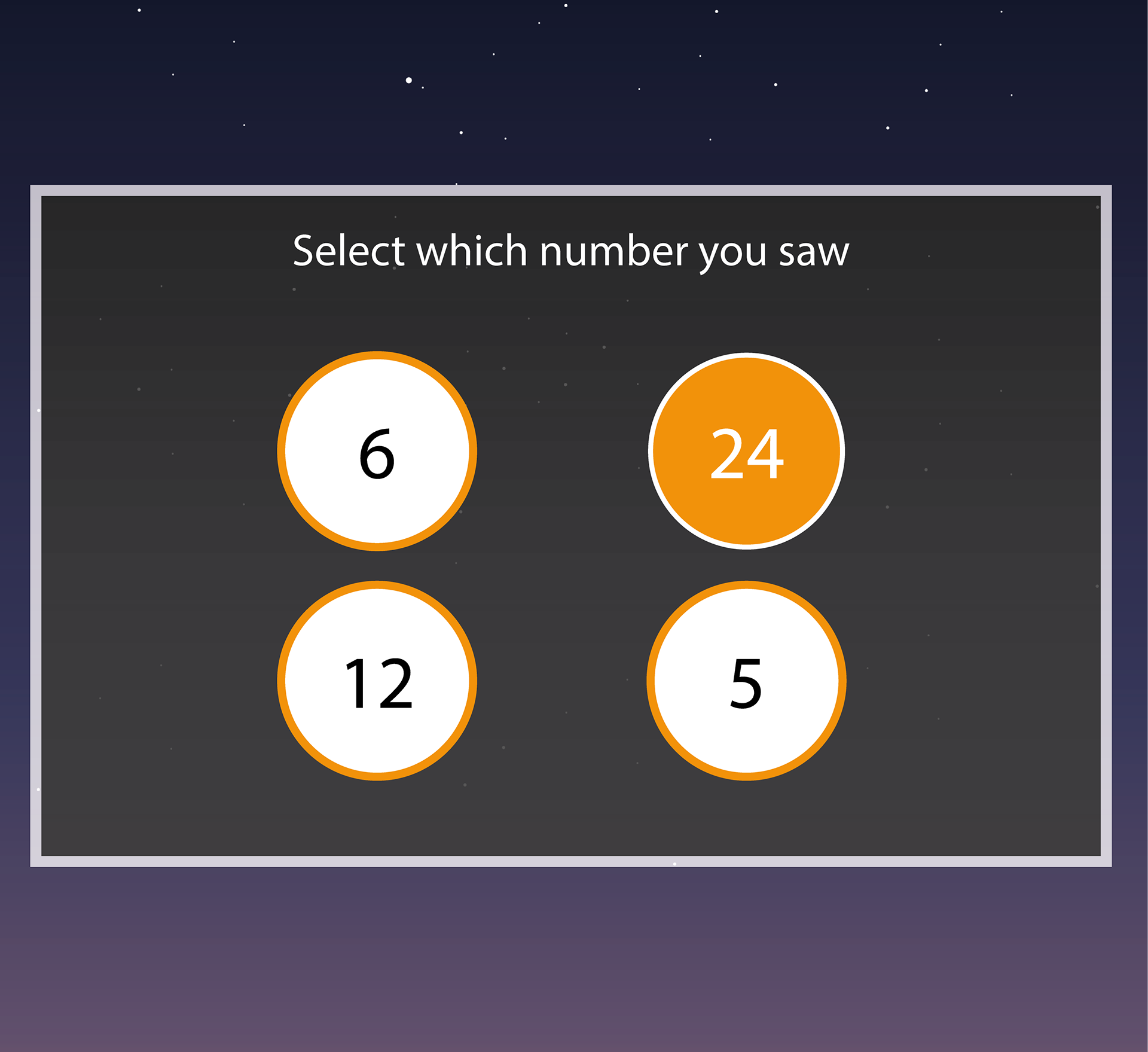
Different screens and tests design for the VR demonstration
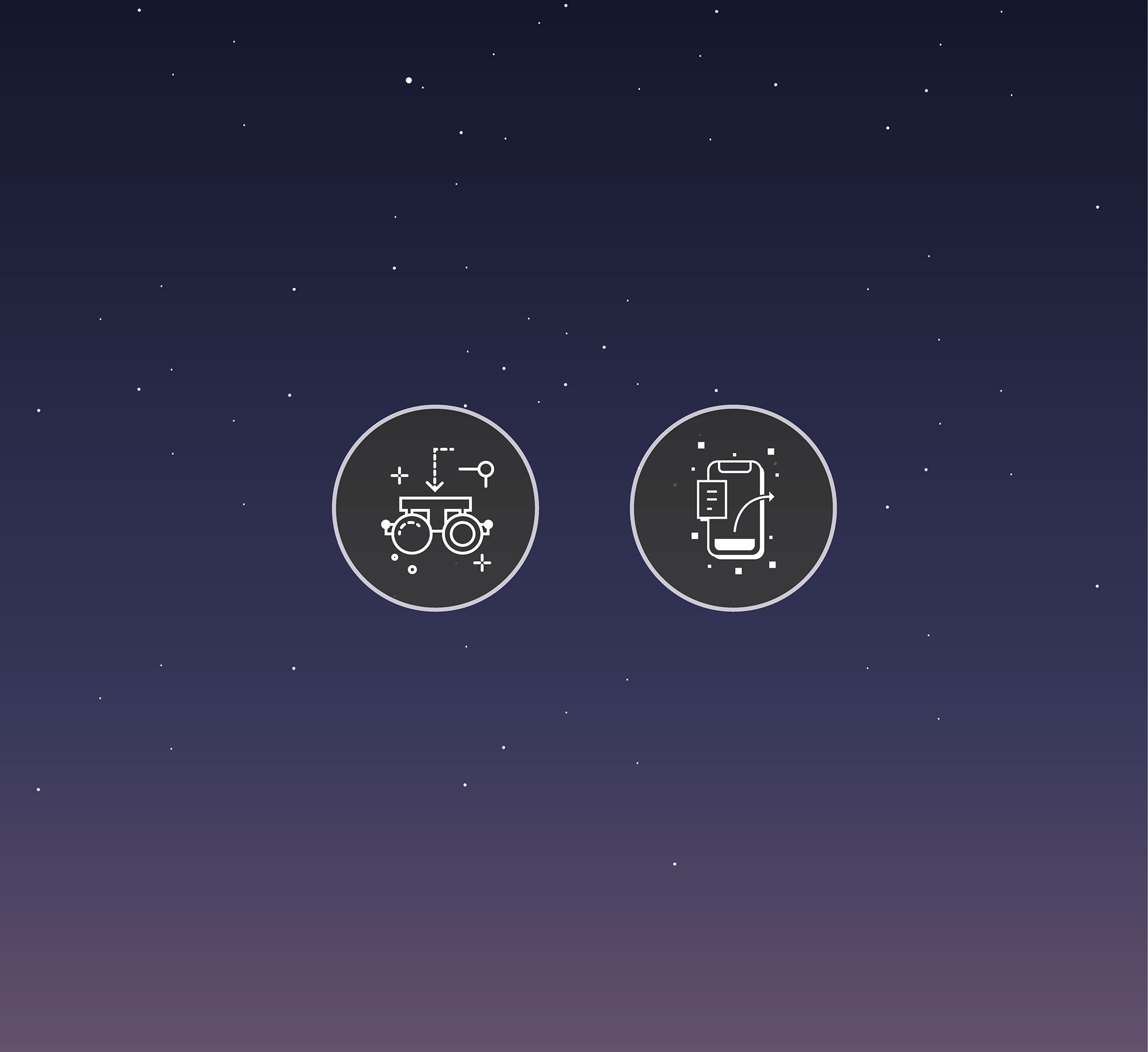
Different screens and tests design for the VR demonstration
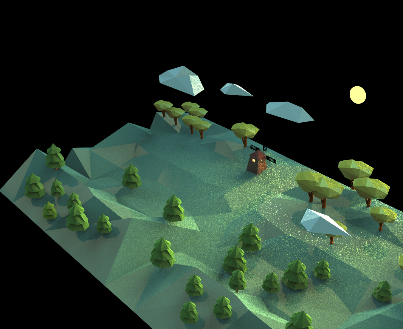
3D model for the VR visual test background. The form is based on the nigh in the forest
The demonstration screens of the low contrast test
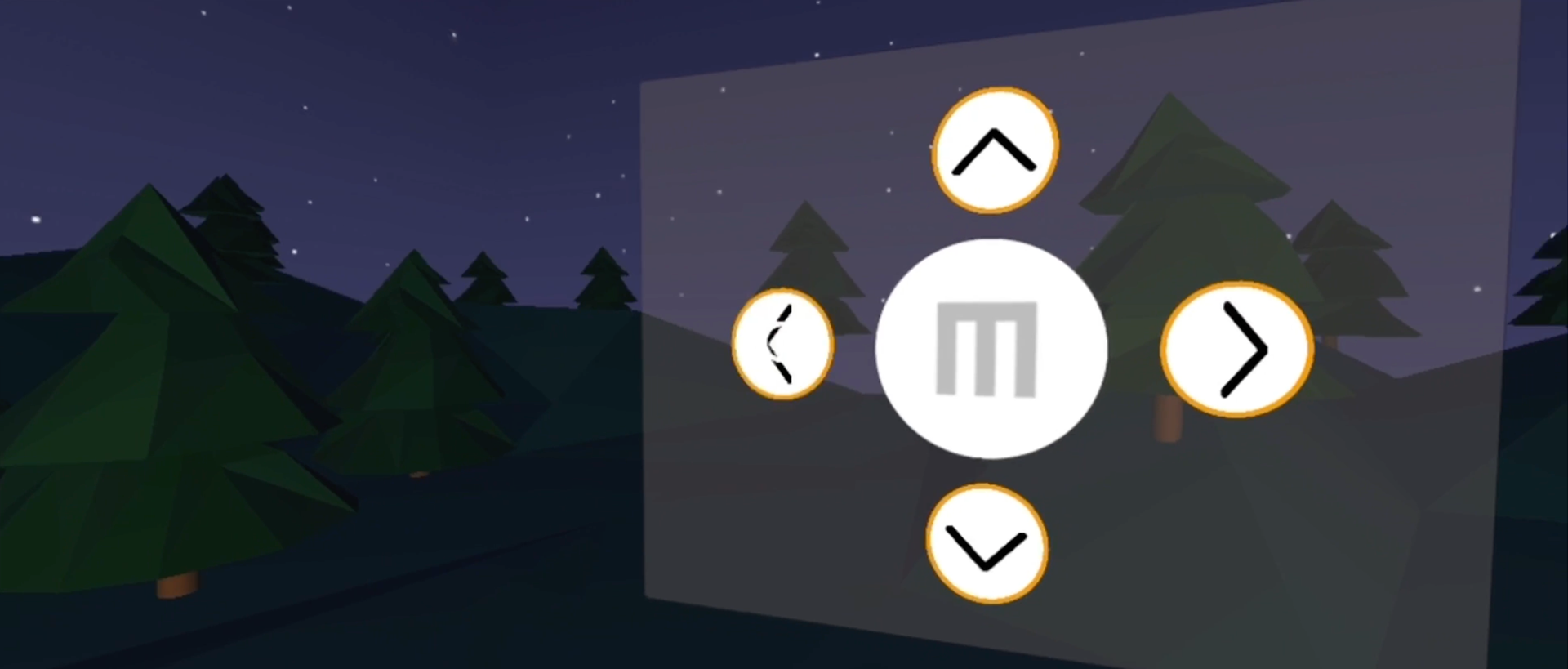
The demonstration screens of the low contrast test
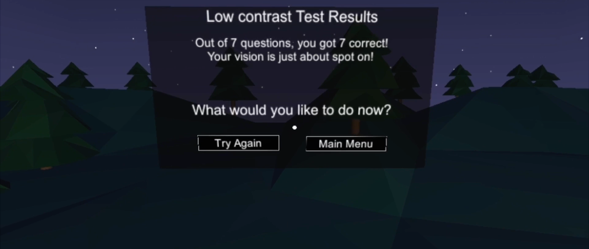
The demonstration screens of the low contrast test
Business plan
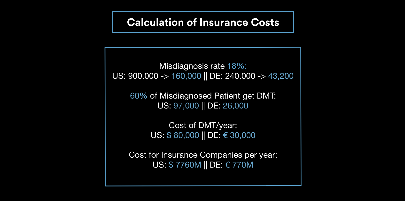
Calculation of insurance costs
Concept presentation video
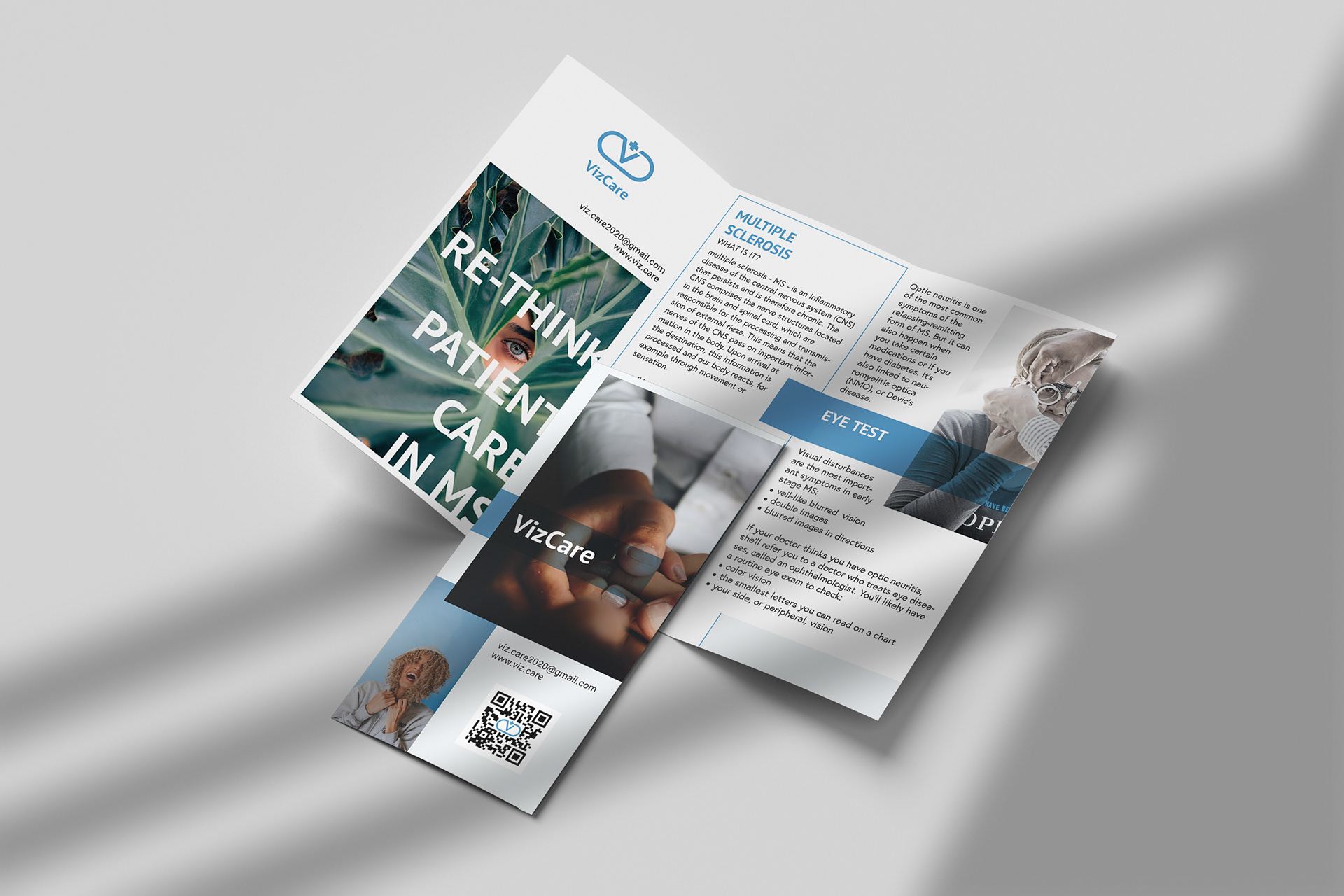
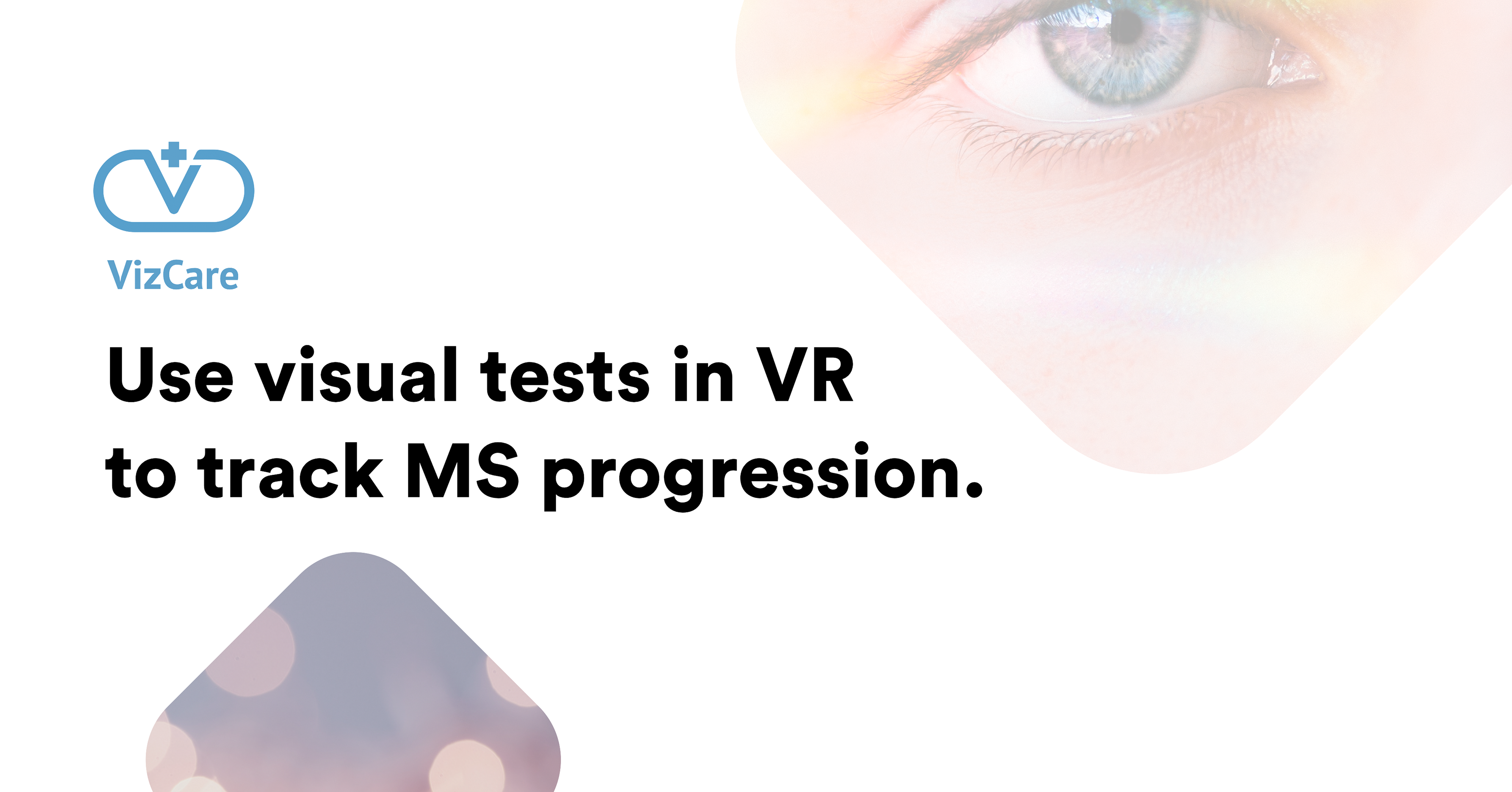
Flyer design
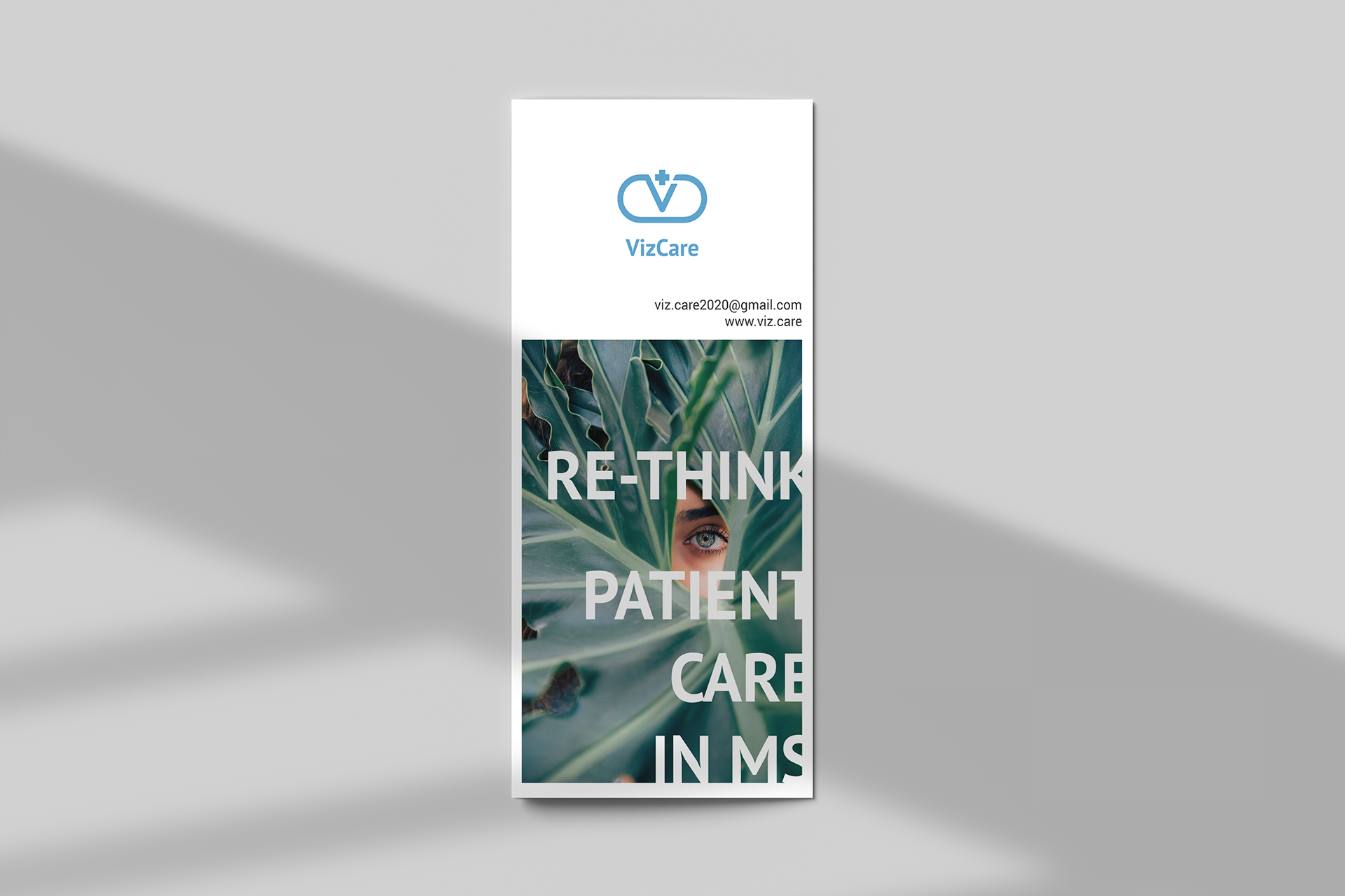
Flyer design
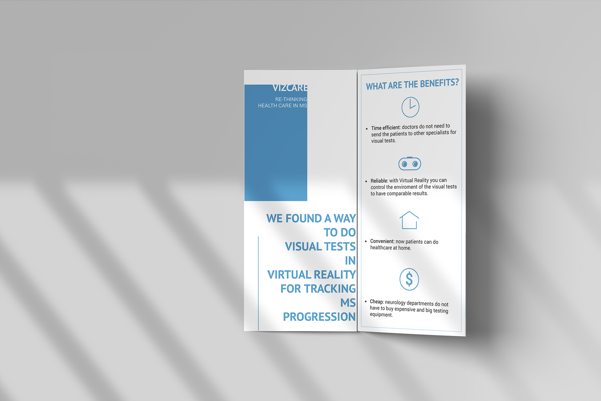
Flyer design
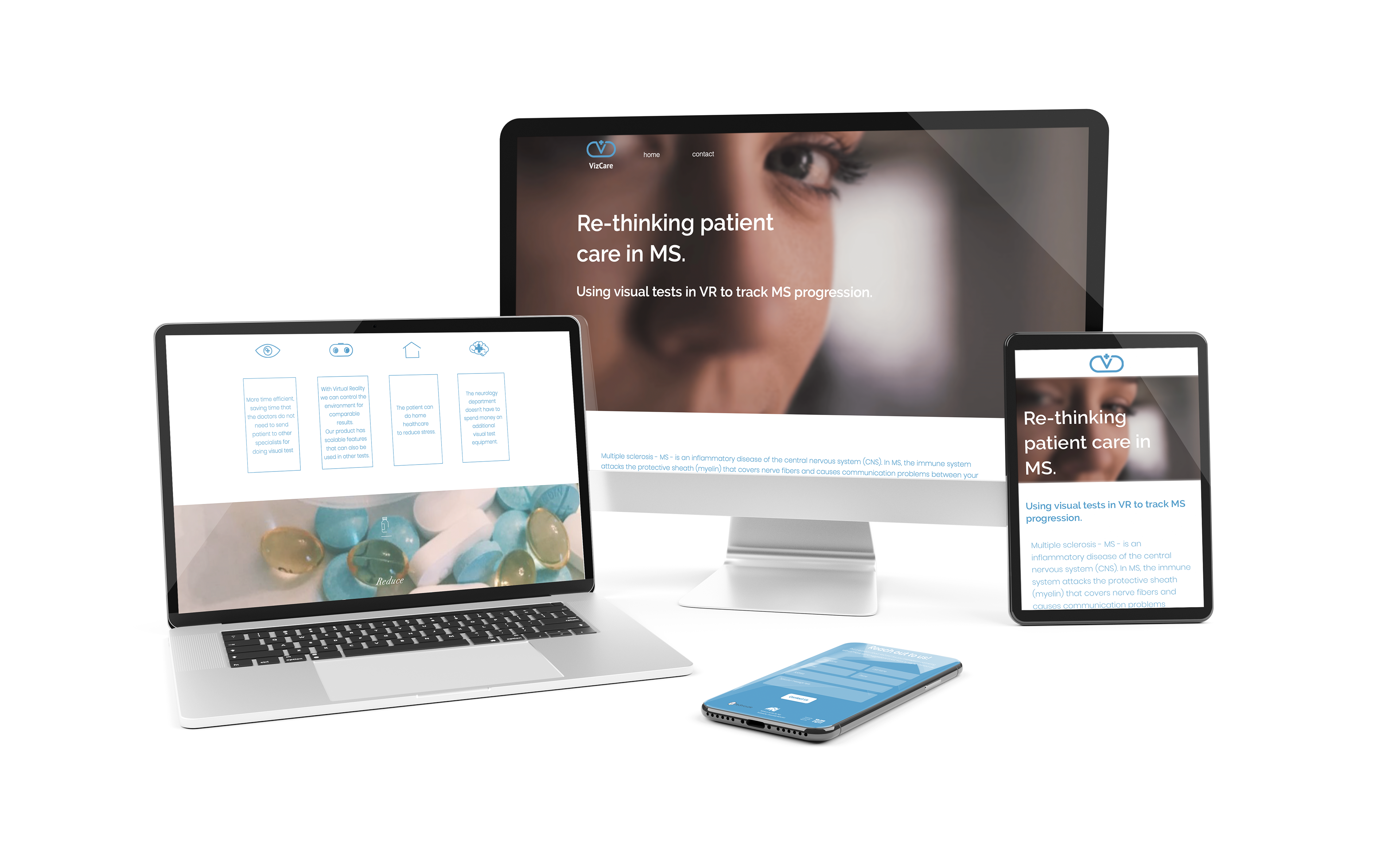
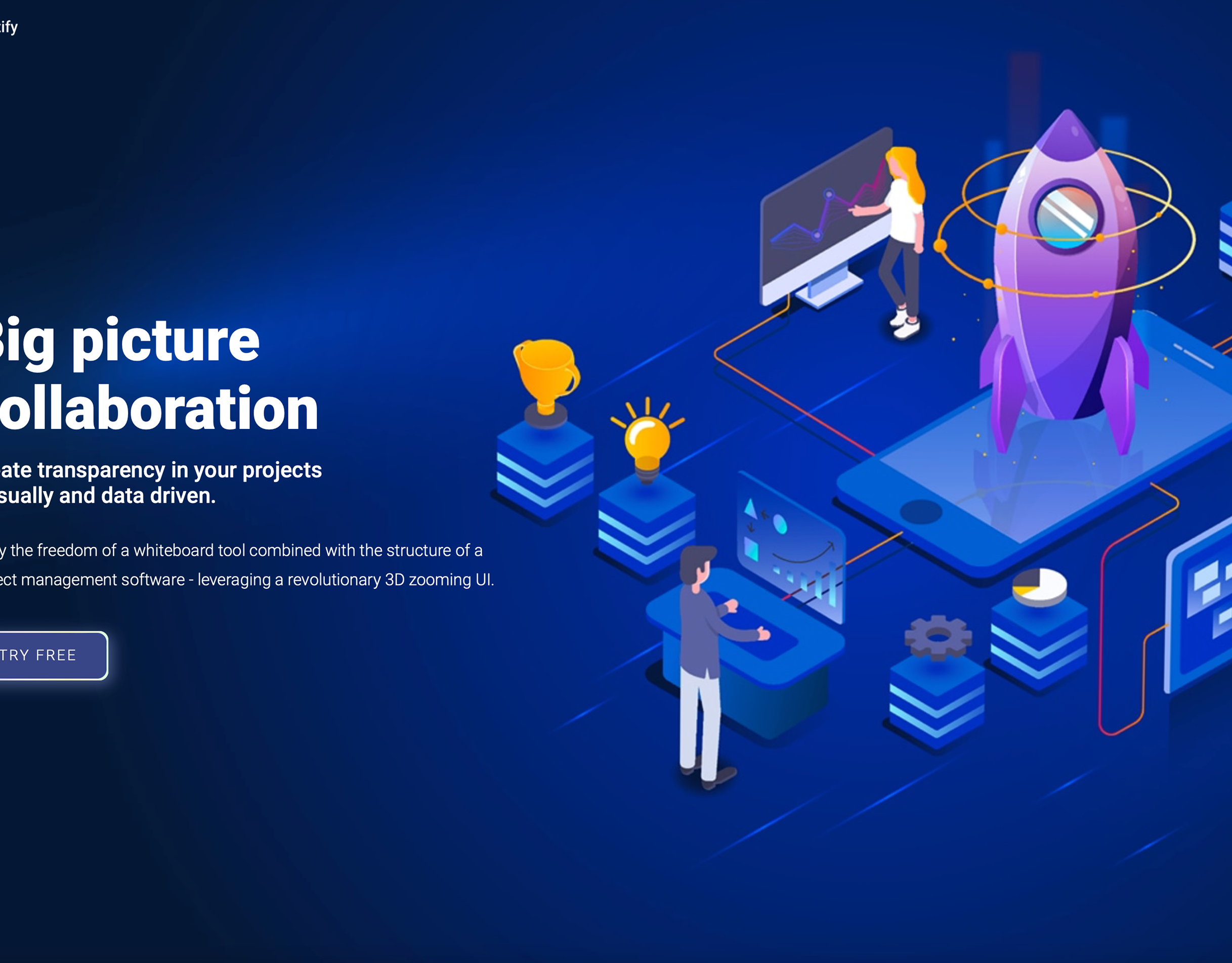
Website design on different devices